
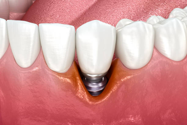
Retrograde peri-implantitis is an uncommon inflammatory condition affecting the bone around the tip, or apex, of a dental implant. Conventional peri-implantitis starts near the gumline and causes progressive bone loss from the top down. Retrograde peri-implantitis typically produces a localized lesion around the implant apex instead.
Possible causes include infection left from a previously extracted tooth or infection from a neighboring tooth. Clinicians also use the terms apical peri-implantitis and implant periapical lesion. These terms overlap, but a dark area at the implant tip on an X-ray does not always mean infection. For patients with dental implants in New London, that distinction shapes every next step.
Identifying the cause takes clinical findings, dental imaging, and an examination of neighboring teeth. Shoreline Periodontics provides this evaluation as part of our dental implant care in New London, CT. Treatment depends on the underlying diagnosis, not on the X-ray appearance alone.
Key Takeaways
- Retrograde peri-implantitis primarily affects the apical portion of a dental implant.
- Marginal peri-implantitis starts around the implant neck and follows a different pattern.
- An old extraction-site infection or an infected neighboring tooth may be associated with an apical implant lesion.
- Symptoms can include pain, swelling, or drainage, but some lesions cause few symptoms.
- A dark area at the implant tip on an X-ray is an important finding, not a complete diagnosis.
- Treatment depends on the cause, implant stability, and whether nearby teeth need root canal care.
What Is Retrograde Peri-Implantitis Around a Dental Implant?
Retrograde peri-implantitis is inflammation in the bone around the deepest part of an implant. It does not necessarily mean that infection began at the gumline and traveled down.
What Does “Retrograde” Mean in Retrograde Peri-Implantitis?
The implant apex is the deepest end of the implant fixture inside the jawbone, opposite the part that holds the crown. “Retrograde” refers to where the problem appears, starting around the apex instead of at the gumline. Clinicians may also describe an implant periapical lesion, a broader term for any abnormality near the tip.
Is Retrograde Peri-Implantitis Common?
No. It is considered uncommon compared with marginal peri-implantitis. Researchers have documented it for decades, including in a 20-year study of retrograde peri-implantitis. Published estimates vary, so no single rate applies to every patient.
Can Retrograde Peri-Implantitis Affect an Implant That Otherwise Looks Healthy?
Yes. The gums and bone near the crown can look normal while a lesion sits around the apex. Healthy-looking gum tissue does not rule out a problem deeper in the bone.
Retrograde Peri-Implantitis vs. Marginal Peri-Implantitis: What Is the Difference?
The two conditions differ mainly in where they appear and what tends to cause them. Retrograde lesions often relate to infection around a tooth root, which explains their position at the apex.
| Characteristic | Retrograde peri-implantitis | Marginal peri-implantitis |
| Primary location | Implant apex | Implant neck, near the gumline |
| Typical bone finding | Localized apical lesion or dark area | Progressive bone loss from the top |
| Frequent clinical context | Previous extraction-site or neighboring tooth infection | Plaque-related inflammation |
| Gumline inflammation | May be absent | Common |
| Bleeding on probing | May be absent | Commonly present |
| Swelling or drainage | May occur near the apical region | May occur around the implant neck |
| Implant stability | May initially be preserved | Often preserved until advanced disease |
| Key diagnostic workup | Apical imaging, implant assessment, neighboring tooth evaluation | Probing, inflammation, and bone-level assessment |
| Treatment considerations | Source-specific endodontic or surgical management | Disease control and ongoing maintenance |
Where Does Marginal Peri-Implantitis Begin?
Marginal peri-implantitis begins in the tissues around the implant neck, near the gumline. Plaque biofilm triggers inflammation, which can lead to progressive loss of crestal bone, the top edge of the bone supporting the implant. Our guide to treatment for marginal peri-implantitis explains how that disease is managed.
Why Bleeding Gums May Be Absent in Retrograde Peri-Implantitis
Periodontal probing measures the tissues around the implant neck. A retrograde lesion may sit much deeper, near the tip. An absence of gumline bleeding does not rule out apical disease.
Can Retrograde and Marginal Peri-Implantitis Occur Together?
Yes. More than one disease process can affect the same implant. A defect that spreads from the gumline toward the tip, however, differs from an isolated apical lesion. Clinicians avoid labeling every lesion that reaches the apex as retrograde peri-implantitis.
What Causes Retrograde Peri-Implantitis?
Most explanations involve infection or inflammation that was present before or near the implant.
Residual Infection From a Previously Extracted Tooth
A tooth removed for infection may have had an inflamed lesion around its root tip, called apical periodontitis. Extraction does not always clear every trace of that process right away. A systematic review of retrograde peri-implantitis concluded that an infectious cause is the most common explanation.
Infection From a Neighboring Tooth With Apical Periodontitis
A natural tooth beside the implant may have infected tissue around its root tip. When an implant sits close to that area, the inflammation can involve the bone near the implant apex.
Retained Root Fragments or Surgical Bone Injury
Occasionally, a small root fragment or foreign material remains at a former extraction site. This does not automatically cause infection. Implant site preparation can also affect surrounding bone. Some of these injuries cause X-ray changes that are not infectious.
Why Retrograde Peri-Implantitis Usually Has More Than One Possible Explanation
Several factors can interact, including pre-existing infection, neighboring tooth disease, local anatomy, surgical history, implant position, and healing. The published evidence comes largely from case reports and observational studies.
Can an Old Root Canal Infection Cause Problems Around a New Dental Implant?
It can in some cases. A past or ongoing root infection may affect the bone at or beside the implant site.
How a Previous Tooth Infection Can Affect the Implant Site
If inflammation from a former tooth’s root persists, it may be associated with a later lesion at the implant apex.
Why an Adjacent Root-Canal-Treated Tooth May Need Evaluation
Root canal treatment is often successful. Still, persistent or recurrent infection around a treated root can occur in some cases, and patients may not know about it. A nearby root-canal-treated tooth may need evaluation if imaging suggests a persistent lesion. An endodontist, a root canal specialist, can assess it.
Why the Neighboring Tooth Is Evaluated Before Removing the Implant
The place where a lesion appears on an X-ray is not always where it started. If a neighboring tooth is the source, treating that tooth may be part of the plan. Locating the source comes before any irreversible decision about the implant.
What Are the Symptoms of Retrograde Peri-Implantitis?
Localized Pain, Pressure, or Tenderness Near the Implant
Some patients notice tenderness, pressure, or discomfort near the implant. Pain alone does not separate apical disease from other early signs of dental implant failure. Avoid pressing on the area repeatedly to test it.
Swelling, a Gum Pimple, or Persistent Drainage
Swelling may appear in the gum near the affected area. Some patients notice a small bump on the gum, called a fistula or draining sinus tract. Pus, drainage, or a recurring bad taste can also suggest an underlying infection. The opening is not always directly over the implant tip, and swelling around a dental implant has several possible causes.
Can Retrograde Peri-Implantitis Occur Without Pain?
Yes. Some apical lesions are found on routine X-rays when the patient reports few or no symptoms.
How Soon After Dental Implant Surgery Can Retrograde Peri-Implantitis Develop?
Timing varies widely. Published case reviews describe diagnoses ranging from about a week to several years after placement.
When Apical Implant Lesions Tend to Appear
Many reported cases appear during early healing, especially when pre-existing infection may be involved. A suspicious area can show up on X-rays before the crown is placed. Later presentations, months or years after surgery, have also been documented. No strict diagnostic window applies.
How Is Normal Postoperative Discomfort Different From a Developing Apical Lesion?
Mild discomfort is expected after implant surgery and usually improves steadily. Pain that persists or worsens, new swelling, drainage, or a concerning X-ray finding should be evaluated. Contact your implant team rather than waiting for a routine visit.
How Is Retrograde Peri-Implantitis Diagnosed by a Periodontist?
Diagnosis combines history, examination, and imaging rather than one symptom or picture. When you see a periodontist in New London, CT, for a suspected apical lesion, the evaluation follows a structured sequence.
Reviewing the Patient’s Implant, Extraction, and Root Canal History
Your clinician will ask about these details.
- When the implant was placed
- Why the original tooth was removed
- Any previously infected teeth nearby
- Root canal treatment on neighboring teeth
- Symptoms after surgery, including swelling or drainage
- Previous dental X-rays
Examining the Gum Tissue, Swelling, and Any Draining Sinus Tract
The clinician looks for redness, swelling, tenderness, and drainage. When a sinus tract is present, the clinician may use exam techniques and imaging to trace where the drainage begins. Never insert anything into the opening yourself.
Measuring Peri-Implant Probing Depths and Checking for Bleeding
Gentle probing shows the health of the tissues around the implant neck. Shallow pockets without bleeding may point toward a pattern other than marginal disease. They do not prove retrograde peri-implantitis on their own.
Checking Whether the Implant Fixture Is Still Stable
The clinician separates true implant movement from a loose crown, screw, or abutment. A localized apical lesion can exist while the implant itself remains stable.
Taking Periapical X-Rays and Comparing Them With Baseline Images
Periapical X-rays are usually the first imaging step. They should show the whole implant, including the apex and neighboring roots. Comparing them with earlier X-rays shows whether a finding is new, stable, or growing.
When Is CBCT Imaging Helpful for an Apical Implant Lesion?
CBCT, a type of 3D scan, may help when standard X-rays are inconclusive or surgery is being planned. Metal implants can create image artifacts, and every scan adds radiation exposure. Not every patient needs CBCT.
Evaluating Neighboring Teeth and Coordinating With an Endodontist
Nearby natural teeth may be checked with cold tests, other pulp tests, tapping, and gentle pressure. Implants have no living pulp, so they cannot be tested the same way. If a tooth shows signs of root infection, the periodontist may coordinate with an endodontist.
The table below shows the possible explanations a clinician weighs during this process.
| Possible finding | Primary location or pattern | Important diagnostic question |
| Retrograde peri-implantitis | Localized around the implant apex | Is there active apical inflammation or infection? |
| Marginal peri-implantitis | Begins near the crestal bone | Are inflammation, probing changes, and progressive bone loss present? |
| Neighboring tooth apical periodontitis | Around the root tip of a natural tooth | Is the neighboring tooth the source? |
| Persistent extraction-site change | Former tooth socket or apical region | Is the finding stable, healing, or progressing? |
| Early failed osseointegration | May involve more of the implant surface | Has stable bone-to-implant contact developed? |
| Noninfectious apical bone change | May appear near the implant tip | Is there a noninfectious or anatomical explanation? |
| Other jaw pathology | Variable | Does the finding need more imaging, referral, or a tissue sample? |
What Does Retrograde Peri-Implantitis Look Like on a Dental X-Ray?
On an X-ray, retrograde peri-implantitis usually appears as a darker area around the implant tip.
What Is an Apical Radiolucency Around a Dental Implant?
A radiolucency is a darker area on an X-ray where less dense material lets more radiation pass through. It does not identify the cause by itself.
How an Apical Lesion Differs From Crestal Bone Loss
An apical lesion sits around the implant tip, often separated from the bone near the gumline. Crestal bone loss starts at the top of the bone and moves downward, as seen with bone loss around dental implants. Whether the darker area connects to the gumline helps clinicians identify the disease pattern.
What a 2D X-Ray Can Miss and What CBCT Can Add
Standard X-rays flatten three-dimensional anatomy into one image. Overlapping structures can hide bone on the cheek and tongue sides. In selected cases, CBCT can show a lesion’s shape, size, and relationship to nearby anatomy.
Does a Dark Area Around an Implant Tip Always Mean Infection?
No. Several conditions can look similar. Clinicians use serial X-rays and the clinical exam to tell them apart.
What Else Can Look Like Retrograde Peri-Implantitis?
Accurate diagnosis protects patients from both unnecessary implant removal and delayed treatment.
Noninfectious Findings, Old Bone Changes, or Imaging Artifacts
Not every apical finding means active infection. Implant site preparation, healed changes from an old tooth infection, or the original bone shape may leave a darker area. Nearby anatomy, such as the sinus in the upper jaw, and image artifacts can also resemble disease.
Inflammation From a Neighboring Natural Tooth
A lesion around a nearby tooth root can overlap the implant on an X-ray. It may be mistaken for a primary implant problem when the tooth is the actual source.
Marginal Peri-Implantitis Extending Toward the Implant Apex
Advanced marginal disease can spread from the gumline down toward the tip. This continuous defect differs from an isolated apical lesion and needs a different approach.
Early Loss of Osseointegration or a Radiolucency Around the Entire Implant
When an implant never integrates with bone, a dark line may surround much of the implant rather than just the tip. This pattern points toward dental implants that fail to fuse with bone, which is a separate problem.
Other Jaw Lesions That Require Further Investigation
An unusual, enlarging, or atypical finding may need more imaging, referral, or a tissue sample. A biopsy is not routine for every apical finding.
What Is the Difference Between Active and Inactive Implant Apical Lesions?
Some apical findings are actively changing, while others remain stable.
Symptomatic and Asymptomatic Apical Lesions
Pain, swelling, drainage, or a lesion that grows on follow-up X-rays raises concern for active disease. Other lesions are found incidentally and cause no symptoms.
Why Symptoms Alone Cannot Establish Disease Activity
A lack of pain does not prove a lesion is harmless. Pain also does not identify its source. Clinicians combine serial X-rays, implant stability, neighboring tooth tests, and exam findings. A single image cannot confirm bacterial activity.
Why There Is No Single Universally Accepted Classification System
Researchers have proposed different ways to classify retrograde peri-implantitis. No single system is universally adopted, and treatment algorithms continue to evolve.
Does Retrograde Peri-Implantitis Mean a Dental Implant Is Failing?
Not always. An apical lesion is a serious finding, but it does not automatically mean the whole implant has failed.
Why the Implant May Remain Integrated Outside the Apical Lesion
The upper and middle parts of an implant can stay bonded to bone while the tip area is affected. Implant stability and the health of the apical tissues are separate findings. True mobility, however, changes the prognosis and treatment options.
Can the Condition Progress and Affect More of the Implant?
It can if the cause is not addressed. Progression is not inevitable, though.
Does an Implant With an Apical Lesion Always Need to Be Removed?
No. The decision depends on the source, symptoms, lesion size, implant stability, and whether treatment is feasible. The factors that determine whether a failing dental implant can be saved apply here as well.
How Is Retrograde Peri-Implantitis Treated After Diagnosis?
Published reports describe several approaches, and no single method is best for every case.
Why Treatment Must Address the Underlying Source
A lesion caused by a neighboring tooth needs a different plan than one starting at the implant site. When a tooth has confirmed endodontic disease, root canal treatment or retreatment may be considered. Treatment of the tooth is based on its own diagnosis.
Surgical Access and Debridement of the Implant Apex
In selected cases, clinicians have reported surgically opening the area to reach the implant tip. The goal is to remove inflamed or infected tissue and clean the affected surface. Surgery is not required in every case.
What Is Implant Apical Resection?
Apical resection involves removing a small portion of the implant tip in selected situations. Published case reports describe it as a specialized approach. It is not a routine step for most implants.
Can Bone Grafting Be Used After Treating an Apical Implant Lesion?
Regenerative treatment may be considered depending on the shape of the defect and the treatment goals. Clinicians sometimes use bone grafting for dental implants in this setting. Grafting does not guarantee complete bone regeneration.
Are Antibiotics Enough to Treat Retrograde Peri-Implantitis?
Antibiotics may be used in selected situations. They are not a standalone cure, because they do not remove the source of infection or the inflamed tissue.
When Is Implant Removal the More Predictable Option?
Removal may be recommended when the implant is truly mobile, damage is extensive, or appropriate treatment has failed. Timely removal may help preserve bone for a future replacement.
Can Retrograde Peri-Implantitis Be Prevented During Dental Implant Planning?
Careful planning reduces risk. Before placement, clinicians commonly review these factors.
- Any existing infection at the planned implant site
- The pulp and root health of neighboring teeth
- Old root canal treatments and previous X-rays
- How well a former extraction site has healed
- Implant position relative to nearby roots and anatomy
Monitoring Healing With Appropriate Follow-Up Imaging
Follow-up X-rays during healing can reveal changes before the final crown is placed.
Why Careful Planning Reduces Risk but Cannot Eliminate Every Complication
No implant procedure is risk-free, even with thorough planning. The goal is to lower risk and catch problems early.
How Is an Implant-Apex Infection Different From Ordinary Gum Disease?
Periodontitis is a chronic infection of the gums and bone that support natural teeth. An implant-apex lesion is different because it usually relates to infection around a tooth root rather than at the gum.
For that reason, general periodontal care, such as scaling and root planing, does not resolve an apical lesion by itself.
When Should You See a Periodontist About Pain or Swelling Near a Dental Implant?
Schedule an evaluation if you notice any of the following.
- Persistent or increasing pain near the implant
- Swelling or a gum bump that keeps returning
- Pus, drainage, or a bad taste that comes back
- New pain after the implant previously felt comfortable
- An unexplained dark area on an implant X-ray
Let your clinician interpret any X-ray finding for you.
When Symptoms Require Urgent Medical Attention
Seek urgent care for rapidly spreading facial swelling with fever or feeling very unwell. Go to an emergency room for difficulty swallowing or breathing. Do not wait for a routine appointment when signs of a spreading infection appear.
How Is Retrograde Peri-Implantitis Monitored After Treatment?
Follow-up continues after symptoms improve.
Comparing Follow-Up X-Rays With the Original Apical Lesion
Consistent follow-up X-rays let the clinician compare the treated area with the original lesion. Visible bone changes can take longer than symptom relief. No specific healing timeline applies to every patient.
Tracking Symptoms, Implant Stability, and Neighboring Teeth
Each visit checks for returning pain, swelling, or drainage. The clinician also confirms implant stability and reassesses nearby teeth. Unresolved root infection in a neighboring tooth may need further treatment.
Regular implant maintenance still matters for long-term gum and bone health. It supports, but does not replace, the follow-up an apical lesion requires.
Frequently Asked Questions About Retrograde Peri-Implantitis
Is retrograde peri-implantitis the same as peri-implantitis?
No. Conventional peri-implantitis starts near the gumline, with inflammation and progressive loss of supporting bone. Retrograde peri-implantitis describes a lesion around the implant tip, often linked to past or nearby root infection. The two can occasionally occur together, but they follow different patterns and need different treatment.
What causes an infection at the tip of a dental implant?
Possible causes include infection left from a previously extracted tooth and infection around a neighboring tooth root. An old tooth infection that never fully resolved may be associated with a later lesion. Often, more than one factor plays a role.
Can a root canal infection spread to a nearby dental implant?
In some cases, a persistent infection around a neighboring tooth root may involve the bone near an implant tip. This does not mean root-canal-treated teeth are generally infected. Many treated teeth stay healthy. When imaging suggests a nearby lesion, the tooth may need an endodontic evaluation.
Is retrograde peri-implantitis painful?
Sometimes. Some patients feel pain, pressure, or tenderness near the implant. Others have few or no symptoms, and the lesion appears only on an X-ray. Pain level does not reliably show how serious the lesion is or where it started.
Can retrograde peri-implantitis occur without bleeding gums?
Yes. Bleeding usually reflects inflammation around the implant neck. A retrograde lesion sits much deeper, near the tip, so the gums may look and feel normal. A healthy gumline does not rule out an apical problem, which is why X-rays of the whole implant matter.
What does an apical radiolucency around a dental implant mean?
It means an X-ray shows a darker area near the implant tip, where bone may be less dense. This finding needs explanation but is not a diagnosis by itself. Infection, healed bone changes, a neighboring tooth lesion, or anatomy can look similar. Clinical findings and earlier X-rays provide context.
Can an implant have an infection at the tip but still be stable?
Yes, in some cases. The upper and middle portions of an implant can stay bonded to bone while the tip area is affected. Stability is a good sign, but it does not rule out an apical lesion. Both findings shape the treatment plan.
When can retrograde peri-implantitis develop after implant surgery?
Timing varies. Many reported cases appear during early healing. Others have been found months or years later. Because no strict window applies, new pain, swelling, or drainage around an implant deserves evaluation regardless of when the implant was placed.
Is CBCT necessary to diagnose an implant-apex lesion?
Not always. Periapical X-rays are usually the first imaging step and often provide enough information. CBCT may help when standard images are unclear or surgery is being planned. Clinicians weigh its added detail against radiation exposure and possible artifacts from the metal implant.
Can retrograde peri-implantitis heal without surgery?
Sometimes. If a neighboring tooth is the source, treating that tooth may resolve the problem without surgery on the implant. Other cases need surgical care at the implant tip. The right approach depends on the cause, symptoms, and the implant’s condition.
Can an implant with an infection at its tip be saved, or does it need to be removed?
Some stable implants can be kept after appropriate treatment of the apical lesion and its source. Removal is not automatic. It becomes more likely when the implant is mobile, damage is extensive, or treatment has not worked.
Can retrograde peri-implantitis return after treatment?
It can, especially if the original source was not fully addressed. A neighboring tooth with persistent infection, for example, may allow the problem to recur. Follow-up X-rays and exams help detect recurrence early. Reporting new swelling, drainage, or pain promptly gives your clinician the most options.
Who can evaluate suspected retrograde peri-implantitis in New London, CT?
A periodontist is well suited to evaluate implant complications, including lesions at the implant tip. Shoreline Periodontics in New London examines the implant, the surrounding bone, and neighboring teeth. We also provide second opinions for implants placed elsewhere.
Get a Dental Implant Infection or Apical Lesion Evaluated in New London, CT
Pain, swelling, drainage, or an unusual finding on an implant X-ray does not always mean the implant must be removed. Inflammation can develop around the implant tip rather than the gumline, which makes an accurate diagnosis especially important.
At Shoreline Periodontics and Dental Implants in New London, CT, patients can get a specialist evaluation for implant concerns. Understanding the implant’s stability, the location of the affected bone, and the health of neighboring teeth helps determine the next step.
Have you been told there is an infection or dark area near your implant tip? Contact our team, especially if swelling, pain, or drainage persists. Schedule an evaluation with a periodontist in New London, CT at our office on Hempstead Street. Shoreline Periodontics also has offices in Middletown, CT, and Westerly, RI.
